Oral cancer is a serious health issue that can affect people of all ages, and early detection is a major factor in improving outcomes. Each year in the United States thousands of new cases are diagnosed, and while advances in treatment have reduced mortality for some types, detecting suspicious changes early still offers the best chance for successful care. A dental office can play a crucial role in recognizing early signs that patients might otherwise miss at home.
Trends in the causes of oral and oropharyngeal cancers have shifted over recent decades. Traditional risk factors like tobacco and heavy alcohol use remain important, but there has also been a meaningful increase in cancers linked to the human papillomavirus (HPV). That change makes routine screening relevant for a broader segment of the population, not only those with long-standing tobacco or alcohol exposure.
Because small lesions and subtle tissue changes can be painless and easily overlooked, regular screenings incorporated into dental exams allow clinicians to compare findings over time. When a dental team is attentive to changes in the mouth, head, and neck region, they can identify abnormalities sooner and guide patients toward prompt evaluation and follow-up care.
The oral cavity includes many different tissues — the lips, tongue, gums, floor of the mouth, inner cheeks, hard palate, and the oropharynx — and cancers can arise in any of these locations. During a screening, clinicians carefully inspect each area for irregularities such as persistent sores, hardened lumps, white or red patches, or areas that bleed without an obvious cause. The throat and tonsillar region are also examined when appropriate, since oropharyngeal cancers can present there.
Symptoms that patients commonly report before an obvious lesion appears include difficulty swallowing, a persistent sore throat, a feeling that something is caught in the throat, changes in voice, unexplained oral numbness, or persistent ear pain without a clear ear problem. While many of these symptoms have noncancerous causes, their persistence beyond a couple of weeks warrants closer attention.
A careful visual and tactile assessment can detect subtle changes in texture, color, or tissue mobility that are not visible in everyday life. Documenting findings and monitoring any evolving signs at regular intervals helps the dental team determine whether an observed abnormality is stable, resolving, or evolving in a way that requires referral for further diagnostic testing.
Screening is beneficial for all patients as part of routine dental care, but certain individuals may require more frequent or vigilant monitoring. People with a history of tobacco or heavy alcohol use, a prior head and neck radiation exposure, occupational chemical exposures, or a history of oral premalignant lesions are typically considered higher risk. Men over 50 have historically shown higher incidence rates, though HPV-related cancers have shifted the risk pattern for some groups.
Because HPV plays a role in many recent cases of oropharyngeal cancer, younger adults without traditional risk factors should not assume they are exempt from screening. Patients who have persistent oral symptoms, noticeable tissue changes, or any new lumps in the neck should be evaluated promptly irrespective of age. The dental team will recommend a screening frequency based on each patient’s medical history and risk profile.
For most patients, an oral cancer screening is integrated into the routine dental exam schedule — typically once or twice a year during regular preventive visits. Patients with higher risk factors or previous suspicious findings may be seen more often so clinicians can track any changes closely and coordinate timely diagnostic steps when needed.
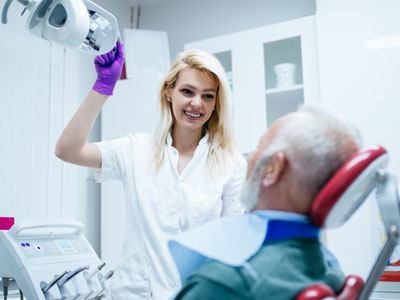
An effective screening begins with a review of the patient’s medical and dental history to identify risk factors and symptoms worth exploring. The clinician then performs a systematic visual inspection of the lips, cheeks, gums, tongue (including underside and sides), floor of the mouth, hard palate, and throat when visible. Good lighting, magnification when needed, and careful technique help reveal subtle findings.
Palpation of the oral tissues and the lymph nodes in the neck is an important component of the exam. Feeling for firm areas, fixed lumps, or enlarged nodes helps the clinician assess whether an area of concern is superficial or may have deeper involvement. If anything atypical is discovered, clinicians will describe the finding, document its appearance and size, and determine the most appropriate next steps.
Adjunctive tools — such as special visualization lights, imaging referrals, or brush biopsies — may be used selectively to gather more information, depending on the nature of the finding. It is important to understand that screening itself is not a definitive diagnosis; rather, it is a way to identify abnormalities that require further evaluation by a specialist or through biopsy to establish a definitive diagnosis and treatment plan.
Prevention and risk reduction focus on modifiable behaviors and staying current with healthcare recommendations. Quitting tobacco and limiting alcohol are two of the most impactful lifestyle changes for lowering oral cancer risk. Protecting the lips from excessive ultraviolet exposure, maintaining a balanced diet rich in fruits and vegetables, and pursuing vaccination against HPV when appropriate are additional measures that reduce overall risk.
If a screening detects a suspicious lesion, the typical pathway involves additional diagnostic steps arranged in coordination with medical specialists. This may include referral to an otolaryngologist (ENT), advanced imaging, or a biopsy performed by a specialist to obtain tissue for laboratory analysis. Early coordination between dental and medical providers helps ensure prompt, organized care when further evaluation is needed.
Emotional and practical support is an important part of the process; patients should expect clear explanations of findings, options for next steps, and guidance about timelines and follow-up. The dental team’s role is to identify potential issues early, communicate concerns compassionately, and help patients navigate referrals and additional care when indicated.
At Strohman Family Dental in Algona, our goal is to make oral cancer screening a routine, reassuring part of preventive care for every patient. If you have questions about what to expect during a screening, risk factors that may apply to you, or next steps after an abnormal finding, please contact us for more information and to discuss your individual needs.
An oral cancer screening is a focused clinical check of the mouth, lips, gums, tongue, floor of the mouth and nearby throat structures to look for early signs of abnormal tissue. The goal of the screening is to identify persistent sores, patches, lumps, or textural changes that may warrant further evaluation. Screenings are performed as part of a comprehensive dental exam and emphasize early detection to improve the chances of successful treatment.
Screening is not a definitive diagnostic test but a method to recognize findings that require additional investigation, such as imaging or biopsy. Results from a screening help clinicians determine whether to monitor a finding over time or refer a patient to a specialist for prompt follow-up. Clear documentation and communication with the patient are essential components of the process.
Routine screenings increase the likelihood of catching abnormal changes at an early, more treatable stage when outcomes are generally better. Small lesions and subtle tissue differences can be painless and easily missed by patients at home, so systematic checks by a trained clinician provide a valuable safety net. Detecting changes early also reduces the time to diagnostic testing and treatment coordination when needed.
Recent shifts in risk patterns, including a rise in HPV-related oropharyngeal cancers, make routine screening relevant for a broader segment of the population. Regular comparison of findings across visits allows the dental team to identify evolving signs rather than relying on a single observation. Early recognition supports timely referrals and interdisciplinary care when appropriate.
Oral cancer screening is recommended for all patients as part of routine dental care, since anyone can develop lesions that deserve attention. Certain individuals are at higher risk and may need more frequent monitoring, including people with a history of tobacco or heavy alcohol use, prior head and neck radiation, occupational chemical exposures, or previous precancerous lesions. Because HPV-related cancers can occur in younger adults without classic risk factors, clinicians do not limit screening by age alone.
The interval for screening is tailored to each patient's medical and dental history; for many patients it is performed during regular preventive visits once or twice a year. Patients with higher risk factors or previous suspicious findings may be asked to return more often so clinicians can track any changes closely. Your dental team, including clinicians at Strohman Family Dental, will recommend a schedule based on your individual needs and risk profile.
Clinicians inspect the lips, cheeks, gums, tongue (including underside and sides), floor of the mouth, hard palate and visible throat for persistent sores, white or red patches, lumps, thickened or hardened areas, and sites that bleed without clear cause. They also palpate the oral tissues and neck to detect firm masses, fixed lumps or enlarged lymph nodes that may indicate deeper involvement. Symptoms patients often report before an obvious lesion appears include difficulty swallowing, persistent sore throat, a feeling of something caught in the throat, or unexplained oral numbness.
Because many common conditions can produce similar symptoms, clinicians evaluate persistence, size, texture and change over time to determine concern. Any finding that does not resolve within a short, defined interval or that appears to be progressing is documented and may prompt further diagnostic steps. Clear documentation supports informed decisions about monitoring versus referral.
An effective screening begins with a review of the patient’s medical and social history to identify risk factors and current symptoms worth exploring. The clinician then performs a systematic visual inspection under good lighting and uses gentle palpation of the oral tissues and neck to check for abnormal texture, firmness or fixed areas. When available and indicated, magnification and adjunctive visualization tools are used to enhance detection of subtle changes.
The clinician documents any findings with descriptive notes and measurements so they can be compared at subsequent visits, and discusses observations with the patient in clear, compassionate terms. If an area appears concerning, the clinician will explain possible next steps, which may include observation, an adjunctive screening tool, referral for imaging or a specialist evaluation. Screening itself does not establish a diagnosis; it guides the need for definitive diagnostic procedures.
Adjunctive tools such as specialized visualization lights, toluidine blue staining, autofluorescence devices or cytology brushes may be used selectively to provide additional information about a suspicious area. These tools can enhance contrast or help identify abnormal tissue patterns that are harder to see under routine lighting, but they do not replace clinical judgment or a biopsy when one is indicated. Clinicians choose adjunctive methods based on the nature of the finding and the best available evidence for their appropriate use.
When an adjunctive device raises concern, it may prompt a more urgent referral for diagnostic imaging or an incisional biopsy by a specialist. Conversely, adjunctive findings that are not concerning can support a conservative watch-and-wait approach with close re-evaluation. Communication about the limits and role of adjunctive testing helps patients understand why further steps may or may not be necessary.
If a suspicious lesion is identified, the dental team will document its appearance, size and location, discuss the finding with the patient and explain recommended next steps for evaluation. Typical next steps include referral to an otolaryngologist or oral and maxillofacial specialist, advanced imaging, or a biopsy to obtain tissue for laboratory analysis. The intent is to obtain a definitive diagnosis quickly so an appropriate treatment plan can be developed if needed.
The dental team often helps coordinate referrals and will communicate findings to the specialist to support timely care, while also providing emotional support and clear information about timelines and expectations. Many suspicious findings turn out to be benign, but prompt evaluation helps rule out or confirm serious disease at the earliest possible stage. Patients should feel empowered to ask questions about the referral process and what the specialist evaluation will involve.
Risk reduction emphasizes modifiable behaviors such as quitting all forms of tobacco, limiting alcohol intake, protecting lips from excessive sun exposure and maintaining a balanced diet rich in fruits and vegetables. Vaccination against human papillomavirus (HPV) according to current public health guidance reduces the risk of HPV-related oropharyngeal cancers and is an important preventive measure when appropriate. Regular dental visits for professional screening and prompt evaluation of persistent oral symptoms are also key preventive actions.
Good oral hygiene, routine professional cleanings, and timely treatment of suspicious oral conditions support overall oral health and may aid in early detection. Discussing individual risks with your dental or medical provider can clarify personalized steps for prevention and surveillance. Staying informed and proactive about symptoms and appointments improves the likelihood of early detection and intervention.
Human papillomavirus, particularly certain high-risk strains, is a recognized cause of many oropharyngeal cancers and has shifted the epidemiology of these cancers in recent decades. HPV-related cancers often arise in the tonsillar and base-of-tongue regions and may present differently than cancers associated with traditional risk factors like tobacco and alcohol. Because HPV can affect younger and otherwise low-risk individuals, awareness and routine screening remain important across a wide age range.
Vaccination against HPV is an effective preventive measure when given according to recommended schedules, and clinicians can discuss vaccine suitability with patients and families. Screening practices do not change based solely on HPV status; clinicians treat persistent symptoms and suspicious findings the same way regardless of suspected etiology. Open communication about HPV and its implications for risk and prevention helps patients make informed decisions.
At Strohman Family Dental in Algona, the approach to oral cancer screening is integrated into routine preventive care and emphasizes careful history-taking, thorough visual inspection and palpation, and clear documentation of findings. The team prioritizes early detection, compassionate communication and coordinated referrals when additional diagnostic steps are needed. Patients can expect screenings during regular exams and guidance tailored to their individual risk factors and concerns.
The practice uses current clinical standards and may employ adjunctive tools selectively when they add useful information to the clinical assessment. If a concerning lesion is found, the dental team assists with timely referral and follow-up to ensure patients receive appropriate specialist evaluation. This collaborative, patient-centered process supports early diagnosis and organized care when further evaluation is required.