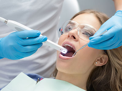
An intraoral camera is a compact, pen-sized imaging tool designed to capture high-resolution, full-color views inside the mouth. Unlike traditional cameras, it’s shaped and sized to glide gently between teeth and along gum lines, producing crisp images of tooth surfaces, restorations, and soft tissues. The live video feed appears on a monitor so both clinician and patient can view the same images in real time, making hidden problems visible without guesswork.
These devices use advanced optics and LED lighting to illuminate hard-to-see areas, which improves contrast and detail compared with unaided visual inspection. Modern intraoral cameras often include autofocus and macro modes that reveal tiny cracks, hairline fractures, or early staining that otherwise might go unnoticed. Because they are digital, the images can be immediately reviewed, annotated, and stored in the patient’s record.
Beyond simple pictures, intraoral cameras are part of a broader shift toward digital dentistry—tools that enhance precision and communication. Their small size and noninvasive nature make them suitable for patients of all ages, and the immediate visual feedback helps clinicians make more confident diagnostic decisions during a routine exam or a focused evaluation.
One of the strongest benefits of intraoral imaging is how it changes the conversation between patient and provider. When a patient can see a clear image of a stained fissure, a chipped edge, or inflamed tissue, abstract descriptions become concrete. This visual context empowers patients to better understand their oral health and to participate actively in decisions about care rather than relying solely on verbal explanations.
Images captured with an intraoral camera can be paused, enlarged, and annotated to highlight areas of concern. Clinicians can point out subtle changes and explain why a condition may need monitoring or treatment. For parents, caregivers, and nervous patients, seeing the issue on-screen often reduces anxiety because it demystifies the problem and the proposed solution.
Because these visuals are preserved in the chart, they also serve as a valuable reference during follow-up visits. Patients can compare “before” and “after” images, which clarifies treatment progress and reinforces preventive advice. That continuity of information strengthens trust and helps patients stay motivated with home care routines recommended by the dental team.
Intraoral cameras assist clinicians in detecting a range of conditions that might otherwise be difficult to catch early. They are particularly useful for identifying fine cracks, early occlusal decay, marginal gaps around restorations, and localized tissue changes. The magnification and lighting reveal surface texture and color shifts that can signal early disease or the need for restorative attention.
Documentation is another critical function. Still images and short video clips become part of the permanent record, supporting accurate charts and longitudinal tracking. These images are invaluable when coordinating care with specialists or dental laboratories, conveying exact details that verbal descriptions may miss. The visual record also supports more efficient case planning and clearer communication across the care team.
Because intraoral photos provide objective evidence of clinical findings, they are often used alongside radiographs and examinations to form a complete clinical picture. When used thoughtfully, they reduce uncertainty during diagnosis and make collaborative treatment planning more precise, whether for restorative needs, periodontal care, or referral management.
Using an intraoral camera is straightforward and comfortable. During your regular checkup or a targeted visit, the clinician or assistant will position the camera in the area of interest and capture several images or a short live video. The device is designed for gentle contact with teeth and gums, and the process takes only a few moments, depending on how many views are needed to document a concern.
As images appear on the monitor, the clinician will review them with you, explaining what each view shows and why it matters. Because the visuals are immediate, discussion and planning happen during the same appointment, allowing decisions about monitoring, preventive measures, or treatment steps to be made with confidence. Patients often appreciate seeing the precise problem rather than relying solely on verbal descriptions.
All images are saved securely in the digital chart so they can be referenced later. That means if a condition needs to be monitored, clinicians can compare subsequent images to assess progression or healing. The efficiency of capturing and storing images also helps streamline visits, keeping the focus on clinical care and patient education.
Intraoral cameras are engineered to integrate cleanly into a practice’s digital workflow. The images they create work alongside digital radiography, digital impressions, and practice management systems to produce a cohesive record. This interoperability speeds diagnosis and treatment planning while maintaining high standards for data security and clinical accuracy.
From a safety and infection-control perspective, the cameras are used with strict protocols. Disposable sleeves or barriers protect the device during use and are changed between patients; the hard components are cleaned and disinfected following manufacturer and regulatory guidance. The noninvasive nature of the camera also means there is no radiation exposure, making it a low-risk adjunct to traditional diagnostic tools.
For clinicians, the value lies in combining visual evidence with clinical experience. For patients, the advantage is clearer understanding and a stronger partnership in maintaining oral health. At Strohman Family Dental, intraoral imaging is one of several technologies used to enhance accuracy and communication, supporting transparent, evidence-based care that helps patients make informed choices.
In summary, intraoral cameras are a practical, patient-friendly imaging tool that improves diagnosis, documentation, and patient engagement. By revealing details that are hard to see with the naked eye, these devices make oral health conversations more precise and actionable. If you’d like to learn more about how intraoral imaging could help during your next visit, please contact us for more information.
An intraoral camera is a small, pen-sized digital device designed to capture high-resolution, full-color images and short video inside the mouth. These cameras use LED illumination and precision optics to reveal tooth surfaces, restorations, and soft tissues with magnification beyond what the naked eye can see. Images are displayed on a monitor in real time so the clinician and patient can view the same visuals together.
Because intraoral cameras are digital, captured photos and clips can be immediately reviewed, annotated, and stored in the patient record. Modern units often include autofocus and macro modes that make it easier to document fine cracks, early staining, or marginal gaps. Their compact, noninvasive design makes them suitable for routine exams and focused evaluations across age groups.
Intraoral cameras enhance diagnostic accuracy by providing magnified, well-lit views of areas that are difficult to inspect visually. The improved contrast and detail help clinicians detect early occlusal decay, hairline fractures, and marginal breakdown around restorations that might otherwise be missed. When combined with tactile examination and radiographs, these images reduce uncertainty and support more informed clinical decisions.
Real-time visualization also allows immediate discussion of findings between the clinician and patient, which accelerates treatment planning when appropriate. The objective nature of photographic evidence aids in tracking changes over time and verifying the effectiveness of prior care. Using images alongside other diagnostic tools creates a more complete clinical picture for personalized treatment recommendations.
The process of using an intraoral camera is quick and comfortable for most patients. During a checkup or targeted assessment, the clinician or assistant will gently place the camera near the area of interest and capture several stills or a short live clip while the images appear on a monitor. The procedure typically adds only a few moments to the appointment and involves light, noninvasive contact with teeth and gums.
As images display, the clinician will review them with the patient and explain what each view shows and why it matters. This immediate feedback helps patients understand clinical findings and participate in decisions about monitoring or treatment. All images are stored in the digital chart so they can be compared at follow-up visits to evaluate progression or healing.
Intraoral cameras are considered low-risk diagnostic tools that do not emit radiation and are safe for patients of all ages. Practices follow strict infection-control protocols by using disposable sleeves or barriers for each patient and by cleaning and disinfecting reusable components according to manufacturer and regulatory guidance. These measures protect both patients and clinical staff while allowing routine use of the device.
Because the cameras contact oral tissues, adherence to sterilization and barrier techniques is essential for safe operation. Documentation of these protocols in office procedures supports consistent compliance and patient safety. When you see intraoral imaging used during an exam, you can expect those standard precautions to be in place.
Images captured with an intraoral camera are saved digitally and become part of the patient’s chart just like radiographs and clinical notes. They can be annotated, timestamped, and organized to support longitudinal tracking of conditions and responses to therapy. Storing images enables efficient comparison of before-and-after views and aids in monitoring areas that require observation over time.
Digital images also facilitate secure communication with specialists or dental laboratories when coordinated care is needed. Because images provide objective visual evidence, they help ensure that referrals and laboratory prescriptions convey accurate clinical detail. Proper integration with practice management systems ensures images remain accessible and protected under the practice's privacy policies.
Intraoral cameras are particularly useful at revealing surface changes such as early occlusal decay, microfractures, and marginal staining that may not be obvious on casual inspection. Magnification and targeted lighting highlight subtle color shifts, surface texture changes, or minor discontinuities in enamel that warrant closer attention. When such signs are detected, cameras help document the finding and guide decisions about monitoring or intervention.
However, visual imaging is only one part of diagnosis and is most effective when used with tactile exams and radiographs. Some internal lesions or sub-surface decay may require X-rays or other diagnostic tests to confirm extent and depth. Thoughtful use of multiple diagnostic tools yields the most accurate assessment of tooth health.
Visual evidence from intraoral cameras turns abstract descriptions into concrete images that patients can understand, improving informed consent and shared decision-making. Clinicians can pause, enlarge, and annotate photos to point out specific concerns, explain the rationale for recommended treatments, and demonstrate expected outcomes. This transparent approach reduces uncertainty and helps patients weigh options based on clear clinical information.
For multidisciplinary cases, stored images streamline collaboration by showing exact surface conditions to specialists and dental laboratories. Accurate visual documentation promotes more precise case planning and minimizes misunderstanding during handoffs. The result is a more coordinated and evidence-based care process for the patient.
Yes, intraoral cameras are generally well suited for pediatric patients and individuals who experience dental anxiety because the device is small, quick, and noninvasive. The immediate visual feedback can help familiarize children or nervous patients with what the clinician is observing, demystifying the exam and building trust. Short capture times and gentle handling make the procedure comfortable for most patients.
Clinicians can use the on-screen images as an educational tool to explain findings in age-appropriate language, which often improves cooperation and understanding. If a patient is especially sensitive, the team can adapt positioning and pacing to maintain comfort while still obtaining useful diagnostic images. The flexibility of the technology makes it a practical option across diverse patient populations.
While intraoral cameras excel at documenting surface detail, they cannot visualize internal tooth structure or bone and therefore do not replace radiographs. Sub-surface decay, root pathology, and some periodontal conditions require X-rays, probing, or other diagnostic modalities to fully characterize the problem. Cameras are a complementary tool that enhance surface assessment but must be interpreted alongside other clinical data.
In addition, limited access or heavy staining can sometimes obscure surface detail and reduce image quality. In such cases clinicians rely on a combination of visual, tactile, and radiographic examination to form a complete diagnosis. Understanding these limitations helps set appropriate expectations for what intraoral imaging can and cannot show.
To learn more about intraoral imaging and how it may be used during your exam, you can call the practice at (515) 295-5200 to ask questions or request that intraoral photography be part of your appointment. The office staff can explain preparation, what to expect during the capture process, and how images will be stored in your digital chart. If you prefer, mention your interest when scheduling so the team can allocate time to review images with you.
The practice location is 301 E Call Street, Algona, AI 50511, and staff will be able to confirm appointment availability and any specific instructions for your visit. During your appointment the clinician will review captured images on-screen and discuss any findings and recommended next steps. Viewing real-time images is an effective way to understand oral health status and participate in care decisions.