Digital radiography replaces traditional film with electronic sensors and computer imaging to capture dental X-rays. Instead of chemical processing and physical film, digital sensors record an image that is immediately available on a computer monitor. This shifts the X-ray workflow from a manual, time-consuming process to a near-instant, highly navigable digital file. For patients and clinicians, that means clearer visual communication and a more efficient visit.
At its core, digital radiography uses the same basic principles as conventional radiography—X-rays pass through tissues and are captured to reveal structure—but improvements in sensor technology and image processing deliver better contrast and detail. These improvements make it easier to identify early signs of decay, bone changes, and other conditions that affect oral health. Because the images are digital, clinicians can manipulate brightness, contrast, and sharpness to optimize visualization without retaking images.
Beyond the technical advantages, the transition to digital imaging reflects a broader move in dentistry toward more precise, patient-centered care. Digital radiography supports faster diagnosis, improved documentation, and closer collaboration among dental professionals. When combined with modern recordkeeping and treatment planning tools, it becomes a foundational technology for high-quality restorative, preventative, and surgical care.
One of the primary advantages of digital radiography is a significant reduction in radiation exposure compared with older film-based systems. Digital sensors are more sensitive to X-rays, so they require less radiation to produce diagnostic-quality images. That reduction is particularly valuable for patients who need recurrent imaging over time, such as those undergoing orthodontic treatment or monitoring chronic conditions.
Comfort and convenience also improve with digital sensors. The compact, thin sensors are designed to fit inside the mouth with minimal discomfort, and images can be reviewed in real time. If an image needs adjustment, the team can correct positioning and recapture quickly, avoiding lengthy retakes. This speed reduces chair time and helps patients better understand their oral health during the same appointment.
In addition to lower exposure and faster capture, digital radiography eliminates chemical processing, which benefits both patient safety and the environment. The move away from film development removes unnecessary handling and potential exposure to hazardous materials, aligning dental practice with modern safety and sustainability standards.
Digital sensors and image software provide clinicians with enhanced diagnostic tools. High-resolution images reveal subtle differences in tooth density, early decay along margins, and fine bone changes that might be harder to detect on film. Clinicians can zoom in, adjust contrast, and compare images side-by-side to improve accuracy when evaluating small or complex problems.
Another advantage is the ability to use image-enhancement features without compromising the original capture. Tools that measure distances, annotate findings, and standardize orientation help clinicians track changes over time and develop precise treatment plans. These capabilities are especially useful when coordinating care across specialties—restorative, periodontal, and oral surgery—so each provider has consistent, high-quality images to inform decisions.
Because digital images integrate with electronic health records, clinicians can rapidly incorporate radiographic findings into a patient’s chart. This streamlined documentation supports clearer treatment explanations and more informed consent, enabling patients to participate actively in decisions about their care.
Digital radiography vastly simplifies image storage and retrieval. Rather than keeping physical film files, a practice can store images securely in a patient’s electronic record, indexed and accessible with a few clicks. This reduces the risk of lost or damaged records and makes longitudinal comparisons—year-to-year or visit-to-visit—quick and reliable for both patients and clinicians.
Sharing images with specialists or transferring records between offices is also faster and more precise. Digital files can be exported or shared securely when coordinating referrals, seeking a second opinion, or planning interdisciplinary care. The ability to send exact images reduces ambiguity and ensures that every provider reviews the same diagnostic information, which improves continuity and outcomes.
Long-term, a robust digital archive supports better preventive care. By comparing archived images, clinicians can spot gradual changes that require intervention before symptoms develop. This archival capability also helps practices maintain thorough records for compliance, quality assurance, and continuity when patients move, change providers, or require future treatment.
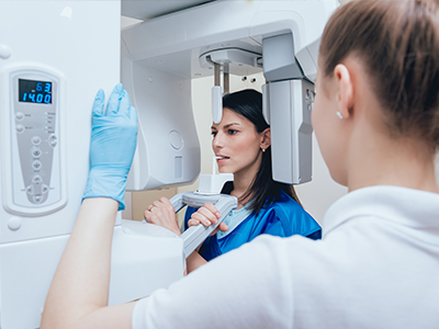
When a patient receives digital X-rays, the process is efficient and patient-focused. The clinician will explain the purpose of the images and position a thin digital sensor in the mouth or align the external unit for panoramic views as needed. Positioning is careful and deliberate to capture the necessary anatomy while maximizing comfort.
Once an image is taken, it appears almost instantly on a monitor in the treatment room. The clinician can review it with the patient, adjust display settings for clarity, and point out areas of interest. This immediate feedback helps patients understand findings and next steps without waiting for film development or external processing.
Because images are captured and stored digitally, follow-up is straightforward. If additional images are required later, the practice can reference prior images for comparison. Patients benefit from clear records and a team that uses modern tools to prioritize accuracy, safety, and clear communication at every visit.
At Strohman Family Dental, digital radiography is part of our commitment to precise, patient-centered care. If you would like to learn more about how we use digital imaging to improve diagnosis and treatment planning, please contact us for more information.
Digital radiography is a method of capturing dental X-ray images using electronic sensors and computer imaging rather than traditional film. The sensors convert X-ray energy into digital files that are available almost instantly for review on a monitor. This immediate availability streamlines workflow and supports clearer visual communication between clinician and patient.
Clinicians use digital radiography because it improves detection of dental conditions and supports more efficient recordkeeping. Digital images can be enhanced to reveal subtle differences in density and structure that may not be obvious on film. These capabilities make digital radiography a foundational tool for preventive, restorative, and surgical decision making.
Unlike film X-rays, digital radiography uses solid-state sensors that record images electronically, eliminating film development and chemical processing. The digital workflow produces diagnostic images with less delay and allows clinicians to adjust brightness, contrast, and magnification without retaking exposures. Sensors tend to be thinner and more durable than film holders, which can improve patient comfort and positioning accuracy.
Digital files are also easier to store, search, and share compared with physical film, which reduces the risk of lost or damaged records. Image enhancement tools help clinicians compare current and prior studies side by side for more consistent evaluations. Overall, the technical and operational differences translate into faster visits and clearer diagnostic information.
Digital dental X-rays use significantly less radiation than older film-based systems because modern sensors are more sensitive and require lower exposure. The radiation dose from routine intraoral digital images is very low, and clinicians follow established guidelines to limit exposure to what is necessary for diagnosis. Protective measures such as lead aprons and thyroid collars are used when indicated to further minimize risk.
Dental teams adhere to the principle of ALARA—"as low as reasonably achievable"—to balance diagnostic benefit with minimal exposure. Patients who require repeated imaging for monitoring receive only the images necessary to manage their care. If you have specific exposure concerns, discuss them with your clinician so they can explain why certain images are recommended.
During a visit that includes digital imaging, the clinician will explain the purpose of the X-rays and position a thin sensor in your mouth or align an external unit for panoramic views. Positioning is done carefully to capture the necessary anatomy while maintaining comfort, and the capture process is quick, often taking only a few seconds per image. If an image needs adjustment, the team can reposition and recapture immediately without long delays for processing.
Once an image is taken it appears almost instantly on the treatment room monitor for review and discussion. At Strohman Family Dental clinicians review images with patients to point out findings and explain recommended next steps using enhanced views when helpful. This immediate review supports clearer communication and informed decision making during the same appointment.
Digital radiography provides high-resolution images and software tools that allow clinicians to zoom, adjust contrast, and measure anatomical features precisely. These enhancements make it easier to identify early decay, assess bone levels, and evaluate root anatomy for endodontic or surgical planning. The ability to compare images side by side aids in detecting subtle changes over time that can influence treatment choices.
Image annotation and measuring tools support accurate treatment planning by documenting dimensions and spatial relationships needed for restorations, implants, and orthodontic assessments. Integration with digital treatment-planning systems and electronic records streamlines coordination across specialties. Together, these capabilities lead to more predictable, personalized care plans.
Digital X-rays are stored electronically in a patient record or secure imaging system, indexed for quick retrieval and long-term archival. Secure storage reduces the risk of lost or damaged film and makes it simple to compare current images with past studies for monitoring. Practices maintain HIPAA-compliant protocols to protect patient privacy when storing and accessing radiographic files.
When coordination with a specialist is needed, digital files can be exported or transmitted securely so every provider reviews the same images. This precise sharing reduces ambiguity and improves continuity of care when multidisciplinary input is required. Electronic transfer also expedites referrals and second opinions without physical media or lengthy courier processes.
Yes, digital radiography enhances the clinician’s ability to identify early signs of disease that are not visible during a clinical exam alone. High-contrast, high-resolution images reveal interproximal decay, small periapical changes, and early bone loss more reliably than visual inspection by itself. Image enhancement tools allow clinicians to adjust visualization to highlight subtle findings that warrant preventive or restorative attention.
Early detection enables interventions that can be less invasive and more conservative, preserving tooth structure and preventing larger problems. By combining radiographic findings with clinical examination, clinicians create a more complete picture of oral health and prioritize timely, targeted care. Regular imaging schedules are tailored to individual risk factors to maximize early detection while minimizing unnecessary exposure.
Digital radiography is suitable for children because sensors require lower exposure and images are captured quickly, which helps minimize motion artifacts and reduces the need for repeat exposures. Pediatric imaging protocols emphasize limiting the number of images to those that are diagnostically necessary and using size-appropriate sensors and positioning aids to maximize comfort. Clinicians also consider caries risk, eruption patterns, and clinical findings when determining the imaging schedule for children.
For pregnant patients, clinicians follow established professional guidance and typically postpone nonurgent radiographs until after pregnancy when feasible. If an X-ray is necessary for urgent diagnosis or treatment, protective shielding and careful exposure techniques are used to minimize any potential risk. Always inform your dental team if you are pregnant so they can adjust care and imaging decisions appropriately.
Digital radiography produces two-dimensional images that are excellent for many diagnostic tasks but have inherent limitations in depicting complex three-dimensional anatomy. Overlapping structures can obscure detail, and certain conditions—such as complex root morphology or evaluation of bony defects—may require three-dimensional imaging like cone beam computed tomography (CBCT). Image artifacts and positioning errors can also affect diagnostic quality and sometimes necessitate repeat or supplemental imaging.
Clinicians determine when advanced imaging is warranted based on the clinical question, treatment complexity, and potential benefit to the patient. CBCT and other modalities are used selectively for implant planning, surgical assessments, and complex endodontic or orthodontic cases where three-dimensional detail improves outcomes. The goal is to choose the imaging approach that provides necessary diagnostic information while minimizing exposure and cost.
Digital radiography enables consistent archival of images that clinicians can compare over months or years to detect gradual changes that signal disease progression. Year-to-year comparisons help identify small shifts in bone level, emerging decay, or changes around restorations that might otherwise go unnoticed. Having a reliable digital archive improves the practice’s ability to intervene earlier with conservative measures when trends indicate increasing risk.
Accessible digital records also support patient education by allowing clinicians to show side-by-side images that illustrate change and explain recommended prevention strategies. At Strohman Family Dental these comparisons help guide personalized recall intervals and prevention plans tailored to each patient’s needs. Well-organized imaging records contribute to continuity of care and more effective long-term oral health maintenance.